How the Lungs Work: Airways, Alveoli and the Muscle That Does the Breathing
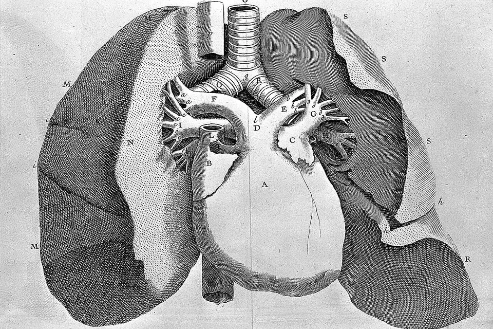
You breathe in and out thousands of times a day without thinking about it. Each breath travels down a branching tree of tubes into hundreds of millions of tiny air sacs, where oxygen crosses into the blood and carbon dioxide crosses out. The lungs themselves have no muscle to do the pumping: that job falls mainly to a thin dome of muscle underneath them, under orders from the brainstem.
Two lungs that are not the same
The lungs fill most of the chest, protected by the rib cage and resting on the diaphragm. They are not a matched pair. According to the US National Heart, Lung, and Blood Institute (NHLBI), the right lung has three lobes (upper, middle and lower) while the left has only two, and the left lung is slightly smaller because the heart takes up room on that side. The left lung even has a notch in its front edge, the cardiac notch, where the heart sits, and a small tongue-shaped extension of its upper lobe, the lingula, takes the place of a middle lobe.
Each lung is wrapped in a two-layered membrane, the pleura. One layer covers the lung, the other lines the inside of the chest wall, and the thin film of fluid between them lets the lungs slide smoothly as they expand and shrink. That fluid also holds the two layers together, a little like two panes of wet glass, which is why the lung follows the chest wall outwards when the chest expands. If air gets between the layers, in a pneumothorax, that grip is lost and the lung can collapse.
The airway tree
Air enters through the nose or mouth, passes the throat and the larynx (voice box), and goes down the trachea, or windpipe, which is held open by C-shaped rings of cartilage. In the chest the trachea divides into two main bronchi, one for each lung. The right main bronchus is wider, shorter and more nearly vertical than the left, which is why an inhaled peanut or small toy more often ends up in the right lung.
The bronchi then divide again and again into ever narrower tubes, the bronchioles. In the classic model published by the Swiss anatomist Ewald Weibel in 1963, the human airway tree branches through about 23 generations between the trachea and the smallest air sacs. Roughly the first 16 generations only conduct air; gas exchange starts in the last few, where the walls of the airways are lined with alveoli.
The conducting airways also clean the air. They are lined with cells that produce mucus and with cells carrying tiny hairs, cilia, which beat in waves to move the mucus, and the dust and microbes trapped in it, up towards the throat, where it is swallowed or coughed out. Physiologists call this the mucociliary escalator. Tobacco smoke damages the cilia, which is one reason smokers cough.
Alveoli: where the exchange happens
The alveoli are tiny, thin-walled air sacs grouped like bunches of grapes at the ends of the airways, each one wrapped in a mesh of capillaries. Their walls and the capillary walls together are extremely thin, so oxygen can diffuse from the air into the blood and carbon dioxide from the blood into the air in a fraction of a second.
How many alveoli are there? The NHLBI says each lung has about 150 million. The most careful count, published by Matthias Ochs and colleagues in 2004, used a stereological method on six adult lungs and found a mean of about 480 million alveoli, with a range from 274 million to 790 million. The number was closely related to the size of the lungs: bigger lungs had more alveoli rather than bigger ones. The honest answer is therefore "several hundred million, and it varies a lot from person to person".
From the alveoli, oxygen-rich blood flows through the pulmonary veins to the left side of the heart, which pumps it round the body. That second, shorter loop of the circulation is described in our page on how the heart works.
The muscle that does the breathing
Lungs cannot inflate themselves. The NHLBI describes the diaphragm, the dome-shaped sheet of muscle separating the chest from the abdomen, as the main muscle of breathing. When it contracts it flattens and moves down, the chest cavity gets larger, the pressure inside the lungs falls slightly below that of the outside air, and air flows in. Breathing out at rest is mostly passive: the diaphragm relaxes and the elastic lungs recoil, like a balloon left open. The intercostal muscles between the ribs help lift and widen the chest, especially during exercise, when the abdominal muscles also push air out actively.
The diaphragm is driven by the phrenic nerves, which leave the spinal cord in the neck, from the third, fourth and fifth cervical segments. Medical students remember it with the rhyme "C3, 4 and 5 keep the diaphragm alive". It explains why a spinal injury high in the neck can stop a person breathing on their own, while an injury lower down usually does not.
At rest, a healthy adult takes about 12 to 20 breaths a minute, the range given in standard clinical references, and each quiet breath moves roughly half a litre of air, a quantity physiologists call the tidal volume. Deep breaths during exercise can move several times as much.
Who decides when you breathe
Breathing is automatic. Groups of neurons in the brainstem, the part of the central nervous system that joins the brain to the spinal cord, set the rhythm and adjust it to the body's needs. The strongest signal is not a lack of oxygen but a rise in carbon dioxide, which makes the blood more acidic; sensors in the brainstem and in the arteries of the neck and chest detect the change and the breathing centres speed up. You can override the system for a while, by holding your breath or talking, but not for long: as carbon dioxide builds up, the urge to breathe becomes irresistible.
Surfactant and the babies it saved
Wet surfaces pull inwards because of surface tension, and an alveolus is a very small wet bubble. In 1929 the Swiss physiologist Kurt von Neergaard showed that surface tension accounts for much of the effort needed to inflate a lung. The lungs counter it with surfactant, a mixture of fats and proteins secreted by cells in the alveolar wall, which lowers surface tension and keeps the smallest alveoli from collapsing at the end of each breath.
In 1959 Mary Ellen Avery and Jere Mead reported that babies who died of hyaline membrane disease, now called neonatal respiratory distress syndrome, lacked surfactant in their lungs. Premature babies often have not yet started producing enough of it. That discovery led, decades later, to surfactant given directly into the lungs of premature newborns, which became standard treatment in neonatal intensive care.
Quick quiz: the lungs
Sources
- National Heart, Lung, and Blood Institute (2022). How the Lungs Work: The Respiratory System and How Your Body Controls Breathing, at nhlbi.nih.gov.
- Ochs, M., Nyengaard, J. R., Jung, A. et al. (2004). The number of alveoli in the human lung. American Journal of Respiratory and Critical Care Medicine 169(1): 120–124, abstract on PubMed.
- Weibel, E. R. (1963). Morphometry of the Human Lung. Springer.
- Avery, M. E. and Mead, J. (1959). Surface properties in relation to atelectasis and hyaline membrane disease. AMA Journal of Diseases of Children 97(5): 517–523.
- Hall, J. E. and Hall, M. E. Guyton and Hall Textbook of Medical Physiology, chapters on pulmonary ventilation and the regulation of respiration. Elsevier.
- Kaiser Permanente Health Encyclopedia, Normal Vital Signs for Adults (resting breathing rate).
 Human Body
How the heart works
Four chambers, four valves, two circuits and the electrical spark behind every beat.
 Human Body
The central nervous system
Brain and spinal cord: how they are built, protected and wired, with the numbers worth knowing.
 Human Body
Bones of the body
Why adults have 206 bones and babies more, and how they split across the skeleton.